alpha300 R – Raman Imaging Microscope
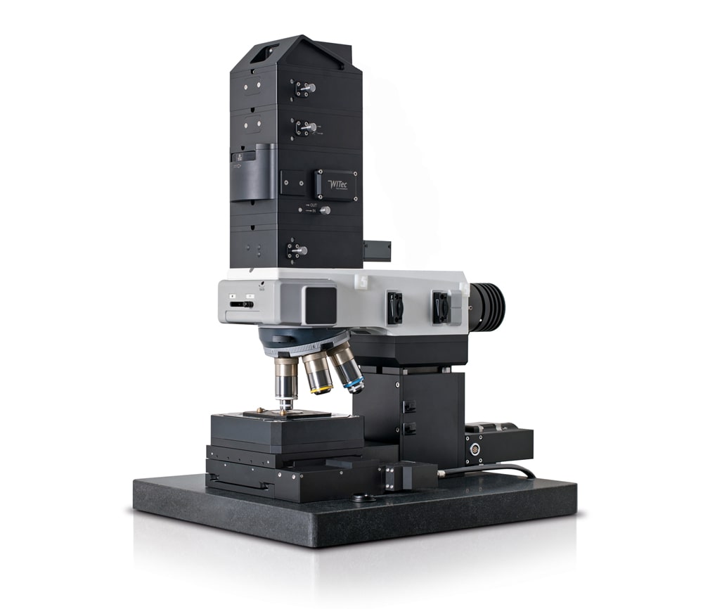
While long recognized as the state-of-the-art imaging system, ongoing development resulting from our innovative spirit keeps the WITec’s Raman microscope alpha300 R at the forefront of the technology and sets the benchmark in terms of flexibility, sensitivity, speed and performance. These unique characteristics have established the alpha300 R the preeminent confocal Raman imaging system on the market.
The flexibility of the alpha300 R series allows the system to adapt to all requirements, combine different imaging techniques and to evolve to meet new or expanded needs.

Key Features
- Confocal Raman Imaging with unprecedented performance in speed, sensitivity, and resolution
- Hyperspectral image generation with the information of a complete Raman spectrum at every image pixel
- Excellent lateral resolution
- Outstanding depth resolution ideally suited for 3D image generation and depth profiles
- Ultra-fast Raman imaging option with under one millisecond integration time per spectrum
- Ultra-high throughput spectroscopic system for highest sensitivity and best performance in spectral resolution
- Non-destructive imaging technique: no staining of fixation of the sample required
Application Examples

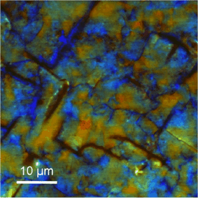
Confocal Raman image of stress in a diamond film. Color coded from red (compressive strain) to blue (tensile strain).

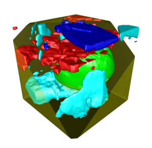
3D Raman Image of a pollen in crystalline honey. Image parameters: 150 x 150 x 50 pixels = 1,125,000 Raman spectra, scan area: 50 µm x 50 µm x 50µm, integration time per spectrum: 12.2 milliseconds, total acquisition time: 3h 50 min.
Color-coded Raman depth-scan of a strcutured GaN layer used for semiconductors. Violet: GaN; Green: fluorescence along the edges of the structure; Red: Stress in material.

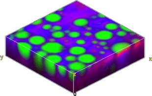
D confocal color-coded Raman image of an emulsion of oil (green), alkane (magenta), and water (blue). 30 µm x 30 µm x 11.5 µm, 150 x 150 x 23 pixels, 517,500 Raman spectra, total acquisition time: 23 min.
Specifications
Raman General Operation Modes
- Raman spectral imaging: acquisition of a complete Raman spectra at every image pixel
- Planar (x-y-direction) and depth scans (z-direction) with manual sample positioning
- Image stacks: 3D confocal Raman imaging
- Time series
- Single point Raman spectrum acquisition
- Single-point depth profiling
- Fibre-coupled UHTS spectrometer specifically designed for Raman microsopy and applications with low light intensities
- Confocal Fluorescence Microscopy
- Bright Field Microscopy
Basic Microscope Features
- Research grade optical microscope with 6x objective turret
- Video system: video CCD camera
- LED white-light source for Köhler illumination
- Manual sample positioning in x- and y-direction
- Fibre coupling
Computer Interface
- WITec software for instrument and measurement control, data evaluation and processing
Raman Optional/Upgradable Operation modes
- Additional lasers, several wavelenghts eligible
- Additional UHTS-spectrometers (UV, VIS, NIR)
- Automated, motorized sample positioning and measuring with piezo-driven scan stages
- Automated confocal Raman imaging
- Automated multi-area and multi-point measurements
- Full automation available: see alpha300 apyron
- Ultrafast Raman imaging, optional available
- Upgradable for epi-fluorescence applications
- Adapter for higher samples
- TrueSurface for Raman depth profiling
- Autofocus
- Dark Field Microscopy, Phase Contrast Microscopy, and DIC optional
Ultrahigh-throughput UHTS Spectrometers
- Various lens-based, excitation optimized spectrometers (UV, VIS or NIR) available, all specifically designed for Raman microsopy and applications with low light intensities
- Fibre-coupled ultrahigh-throughput optical instruments
- Superior peak shape conservation
